Introduction
This tutorial is set to introduce students to the common characteristics of the flowers specimen. The learners will observe the specimen structures from the microscope of the 12 different slides which includes Sunflower Pollen, Bamboo cane, Tulip Pollen and many more.
Setting up the Microscope and slides
- Carefully unpack the microscope from the box. Then place it on a table with an even base.
- Connect the USB cable of the microscope to your laptop.
- Open camera in the laptop and select the camera option on the top left (In my laptop there is a camera rotation option after I have connected the microscope successfully.)
- Now, place a white sheet of paper under the microscope and adjust the focus of microscope till you get a clear image of the white sheet of paper. (Sometime there will be the problem with setting up of the camera try replugging the microscope USB.)
- Carefully place the slide under the microscope and change the focus of the microscope till you get the clear zoomed image of the slide.
Output Flowers specimen
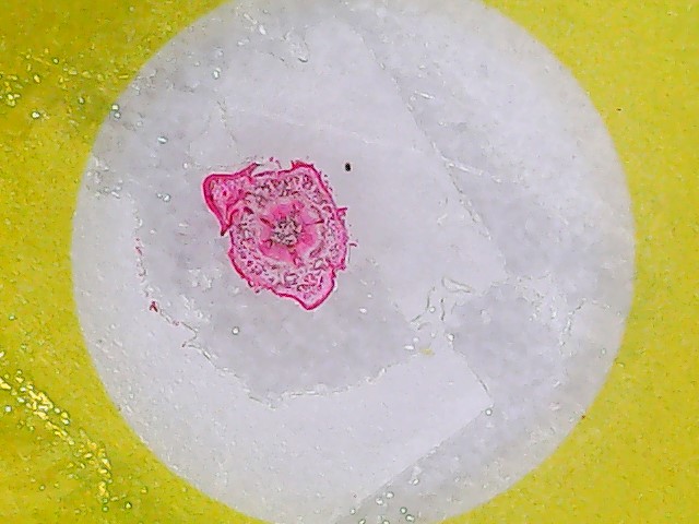
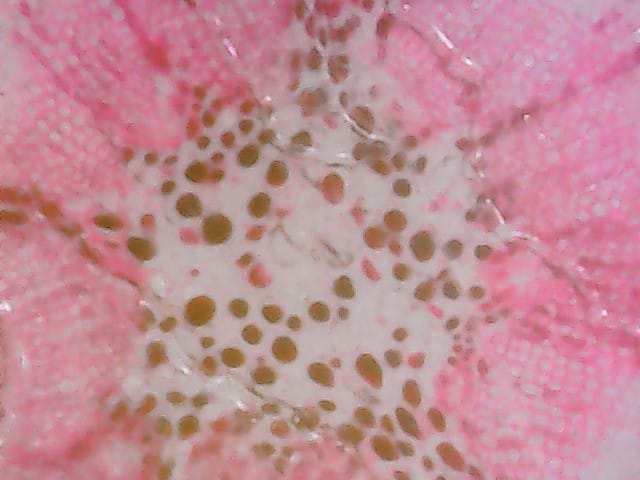
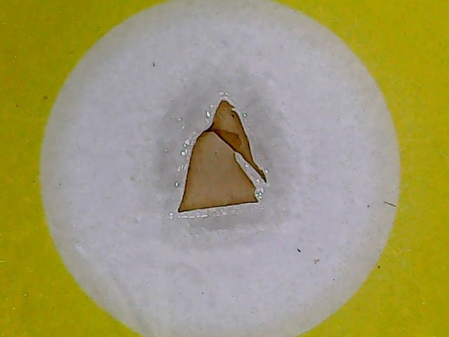
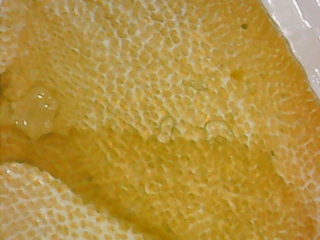
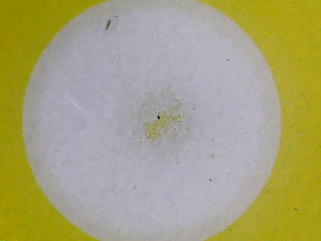
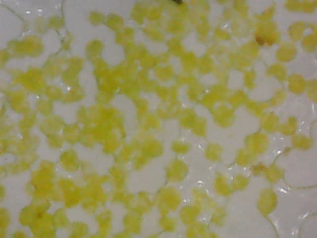
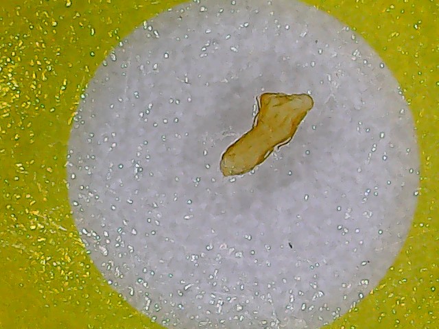
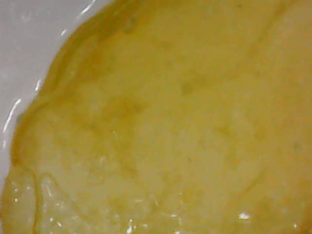
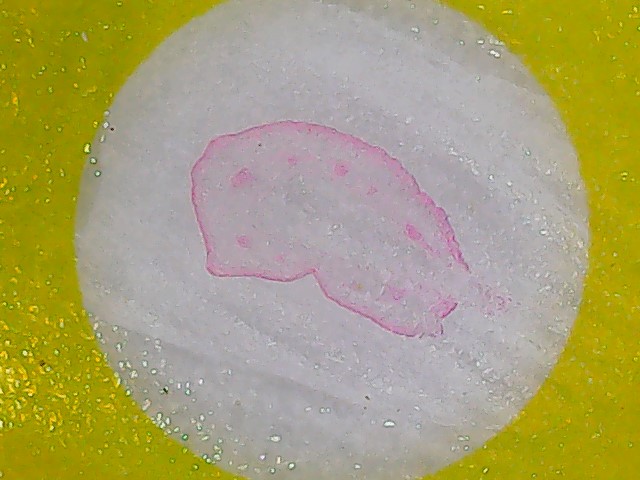
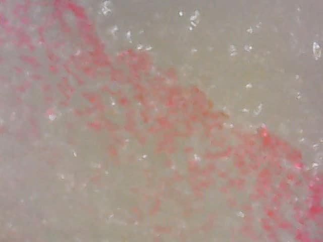
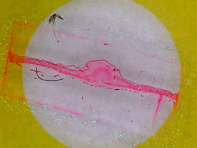
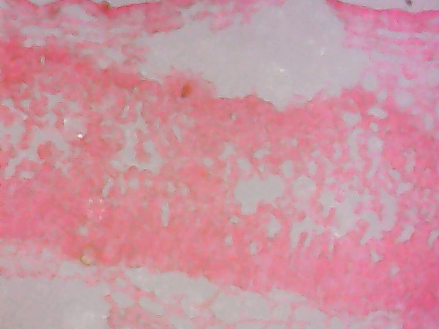
The output images of the slides are shown below with the respective slide names.

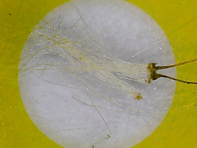
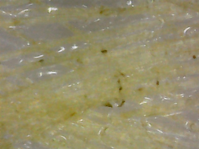
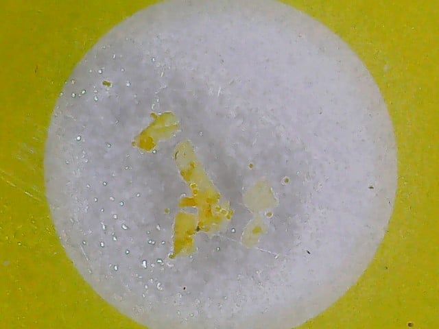
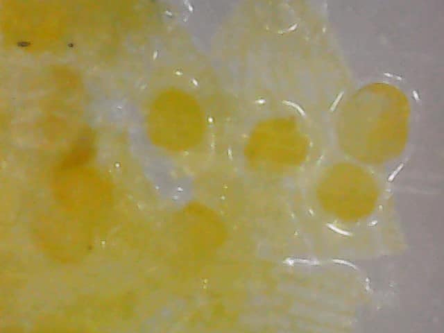
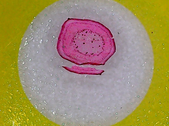
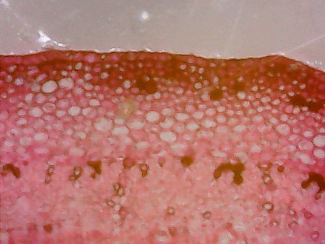
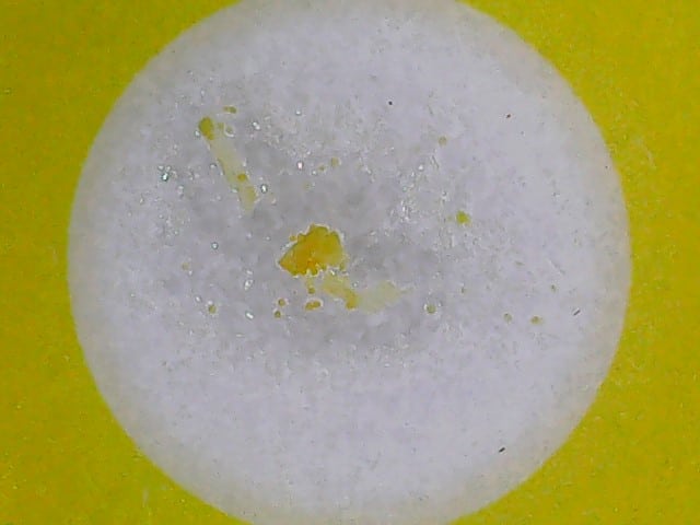
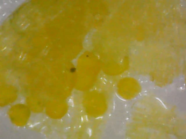
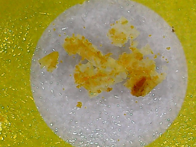
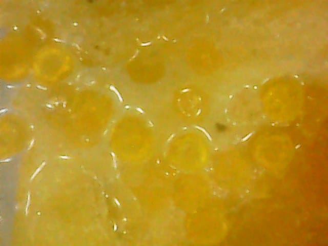
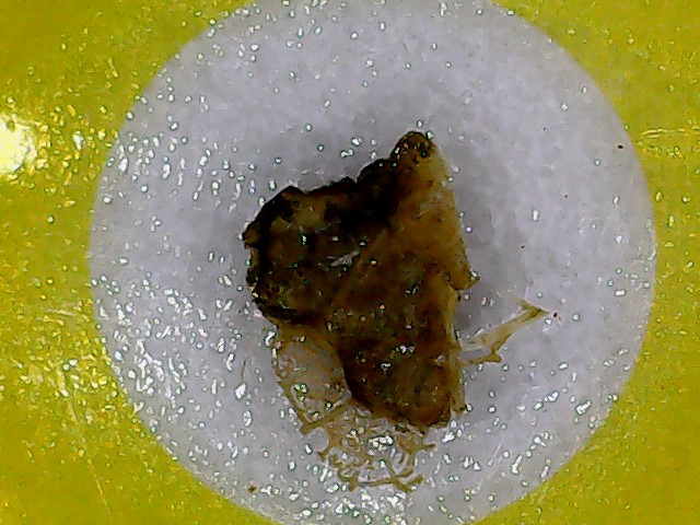
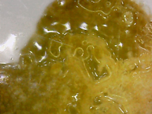
Note: The images on the left are 25 X focused and the one on the right is 800 X focused.
- Laver: This slide sample shows the focused image of Laver using the microscope.
- Sunflower Pollen: This slide sample shows the focused image of Sunflower Pollen using the microscope.
- Agar: This slide sample shows the focused image of Agar using the microscope.
- Bamboo Cane: This slide sample shows the focused image of Bamboo Cane using the microscope.
- Phlox Leaf: This slide sample shows the focused image of Phlox Leaf using the microscope.
- Dandelion fuzz: This slide sample shows the focused image of Dandelion fuzz using the microscope.
- Lily Pollen: This slide sample shows the focused image of Lily Pollen using the microscope.
- Carnation Stem: This slide sample shows the focused image of Carnation Stem using the microscope.
- Camellia Pollen: This slide sample shows the focused image of Camellia Pollen using the microscope.
- Tulip Pollen: This slide sample shows the focused image of Tulip Pollen using the microscope.
- Veins of Holly Leaf: This slide sample shows the focused image of Veins of Holly Leaf using the microscope.
- Pine tree Stem: This slide sample shows the focused image of Pine tree Stem using the microscope.