Introduction
This tutorial is set to introduce students to the common characteristics of the insects specimen. The learners will observe the specimen structures from the microscope of the 12 different slides which includes Locust Antenna, Butterfly Leg, Honeybee wing and many more.
Setting up the Microscope and slides
- Carefully unpack the microscope from the box . Then place it on a table with an even base.
- Connect the USB cable of the microscope to your laptop.
- Open camera in the laptop and select the camera option on the top left (In my laptop there is a camera rotation option after I have connectedthe microscope successfully.)
- Now, place a white sheet of paper under the microscope and adjust the focus of microscope till you get a clear image of the white sheet of paper.(Sometime there will be problem with setting up of the camera try replugging the microscope USB.)
- Carefully place the slide under the microscope and change the focus of the microscope till you get the clear zoomed image of the slide.
Output Insects specimen
The output images of the slides are shown below with the respective slide names.

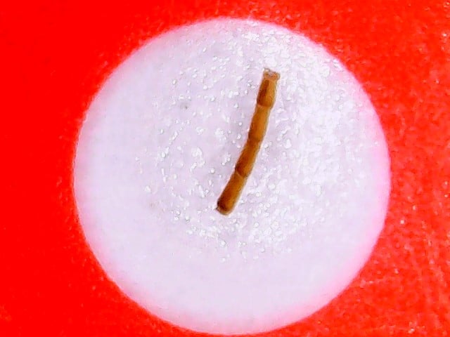
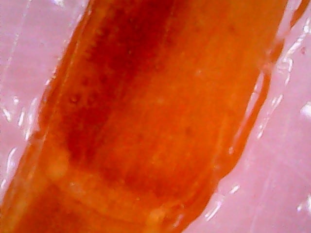
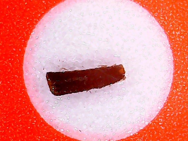
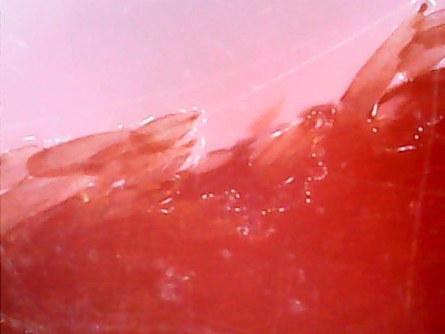
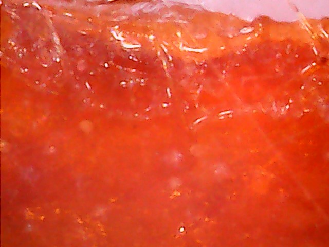
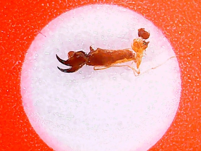
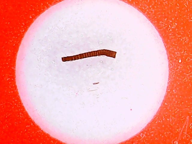
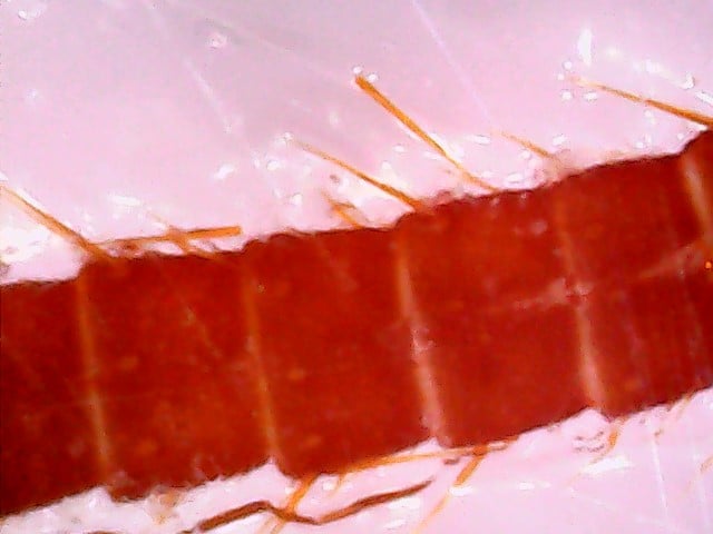
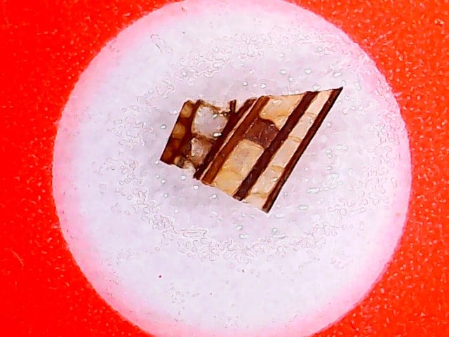
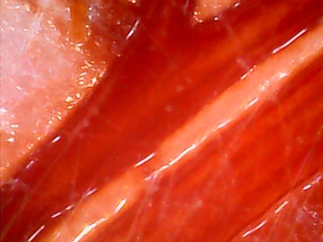
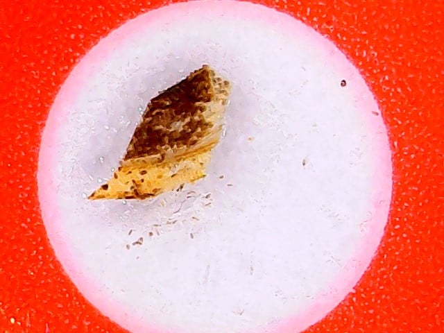
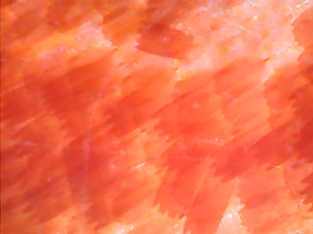
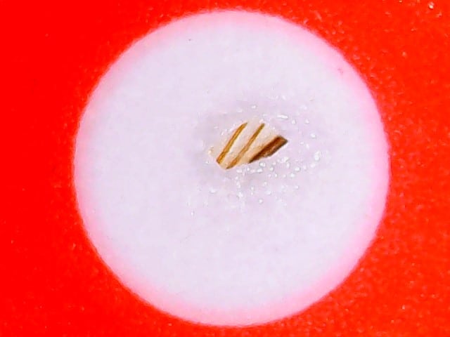
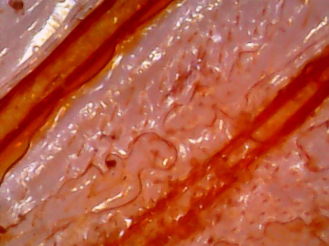
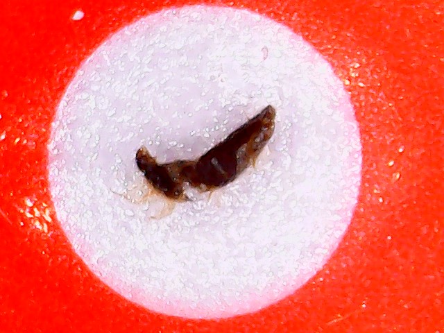
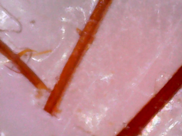
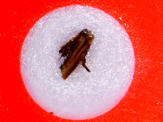
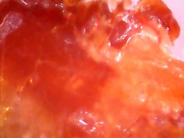
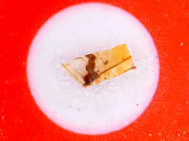
Note: The images on the left are 25 X focused and the one on the right is 800 X focused.
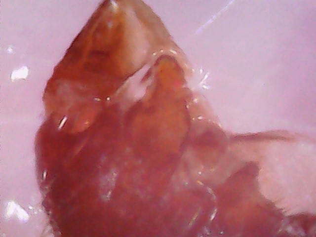
- Locust Antenna: This slide sample shows the focused image of Locust Antenna using the microscope.
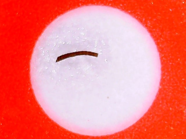
- Butterfly Leg: This slide sample shows the focussed image of Butterfly Leg using microscope.
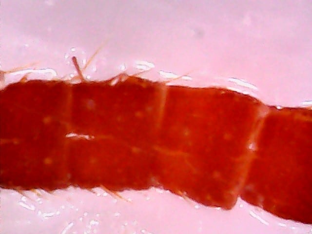
- Locust Leg: This slide sample shows the focused image of Locust Leg using the microscope.
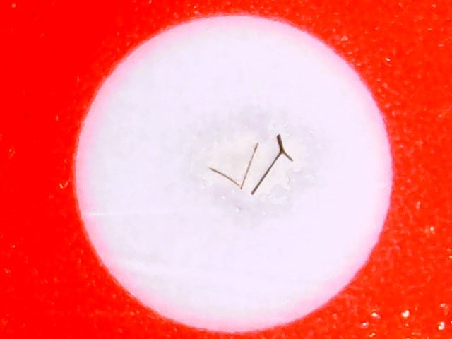
- Butterfly Antenna: This slide sample shows the focused image of Butterfly Antenna using the microscope.
- Locust Wing: This slide sample shows the focused image of Locust Wing using the microscope.
- Butterfly Wing: This slide sample shows the focused image of Butterfly Wing using the microscope.
- Honeybee Wing: This slide sample shows the focused image of Honeybee Wing using the microscope.
- Honeybee leg: This slide sample shows the focused image of Honeybee leg using the microscope.
- Honeybee Antenna: This slide sample shows the focused image of Honeybee Antenna using the microscope.
- Dragonfly Wing: This slide sample shows the focused image of Dragonfly Wing using the microscope.
- Dragonfly Leg: This slide sample shows the focused image of Dragonfly Leg using the microscope.
- Dragonfly Abdomen: This slide sample shows the focused image of Dragonfly Abdomen using the microscope.

Alert: Please handle the slides with care. While placing the slides make sure that you don’t touch the specimen area with your bare hands.