Introduction
This tutorial is set to introduce students to the common characteristics of the Plants Specimen. The learners will observe the specimen structures from the microscope of the 12 different slides which includes Potato starch, Pumpkin Ovary, Onion epidermis and many more.
Setting up the Microscope and slides
- Carefully unpack the microscope from the box. Then place it on a table with an even base.
- Connect the USB cable of the microscope to your laptop.
- Open camera in the laptop and select the camera option on the top left (In my laptop there is a camera rotation option after I have connected the microscope successfully.)
- Now, place a white sheet of paper under the microscope and adjust the focus of microscope till you get a clear image of the white sheet of paper. (Sometime there will be the problem with setting up of the camera try replugging the microscope USB.)
- Carefully place the slide under the microscope and change the focus of the microscope till you get the clear zoomed image of the slide.
Output Plants specimen
The output images of the slides are shown below with the respective slide names.

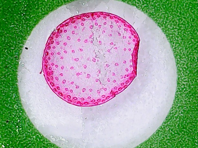
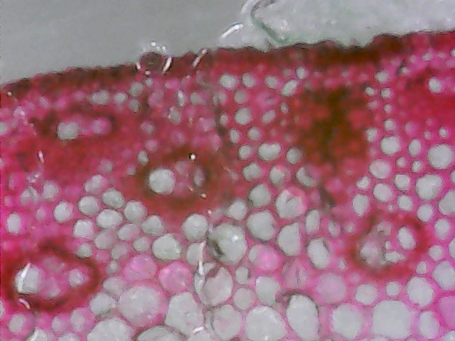
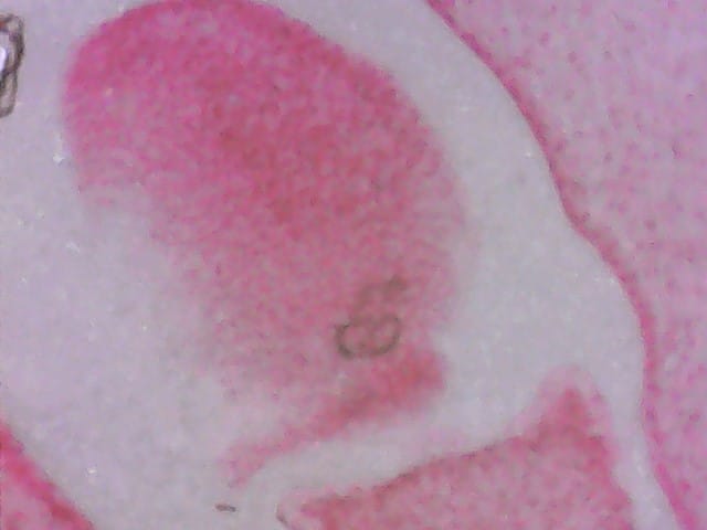
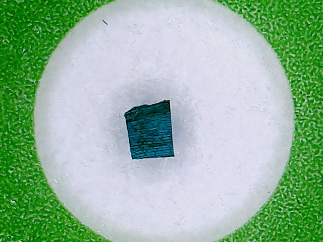
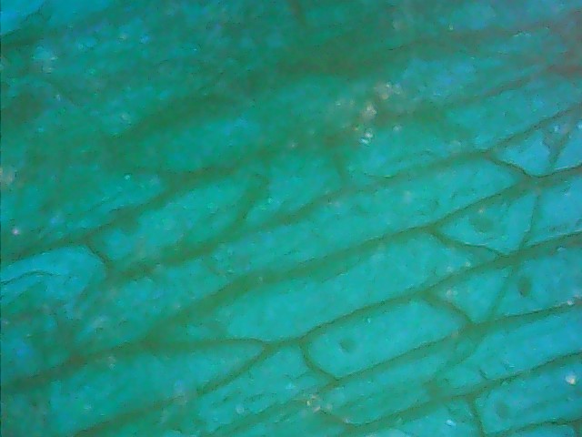
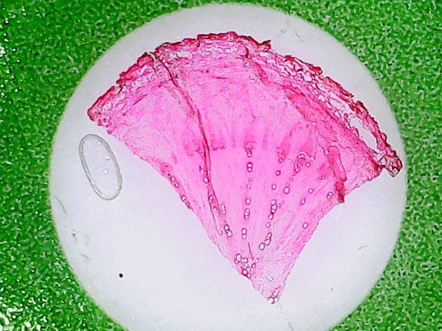
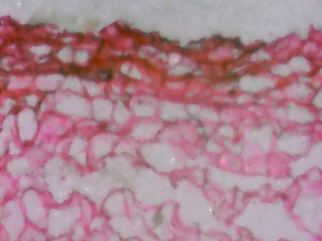
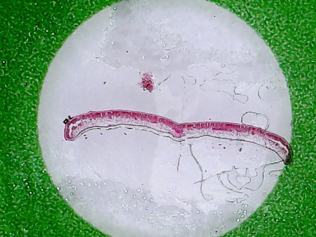
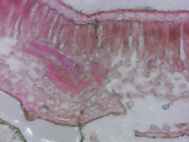
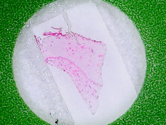
Note: The images on the left are 25 X focused and the one on the right is 800 X focused.
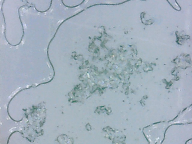
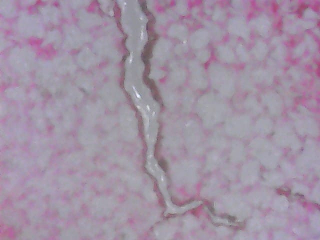
- Potato Starch: This slide sample shows the focused image of Potato Starch using the microscope.
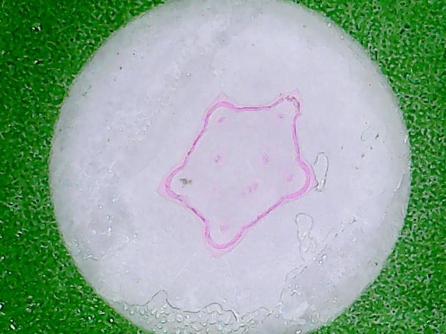
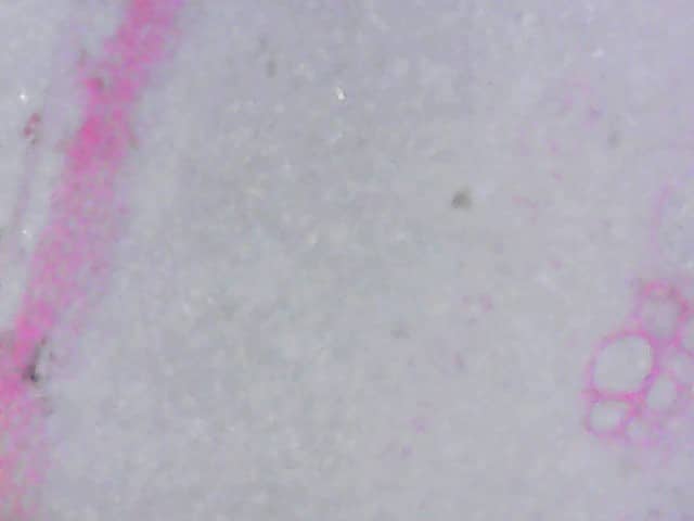
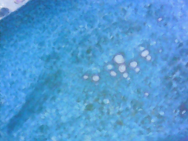
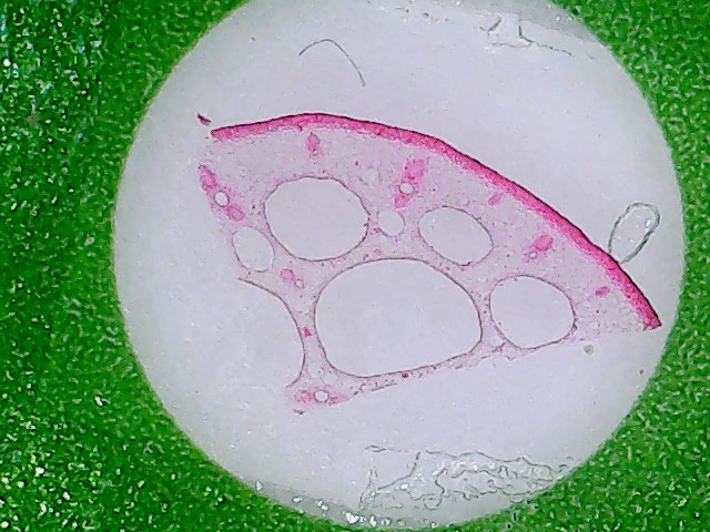
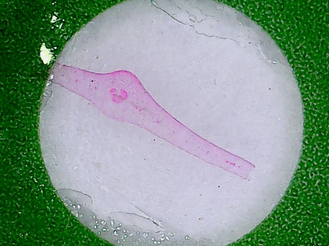
- Sponge Gourd Stem: This slide sample shows the focused image of Sponge Gourd Stem using the microscope.
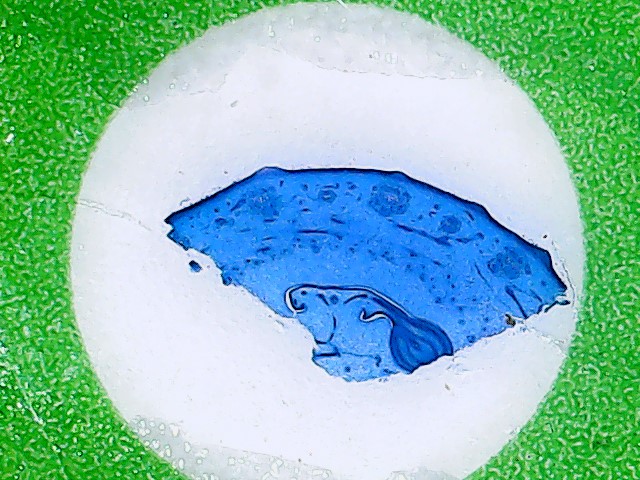
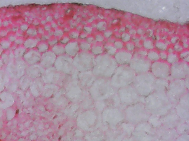
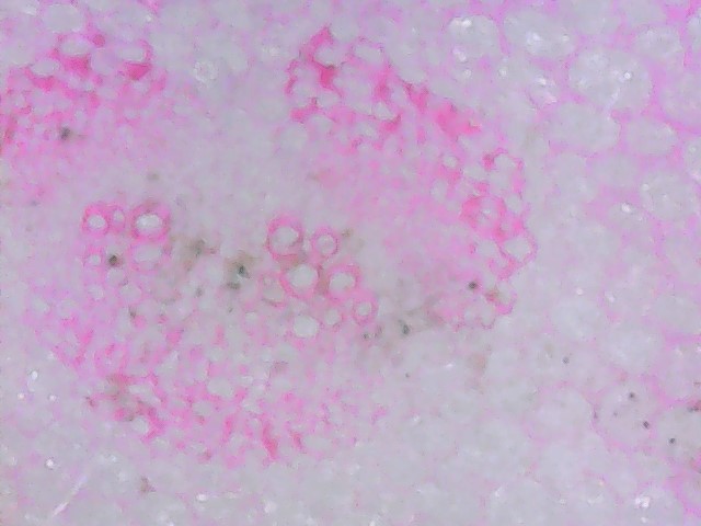
- Pumpkin Ovary: This slide sample shows the focused image of Pumpkin Ovary using the microscope.
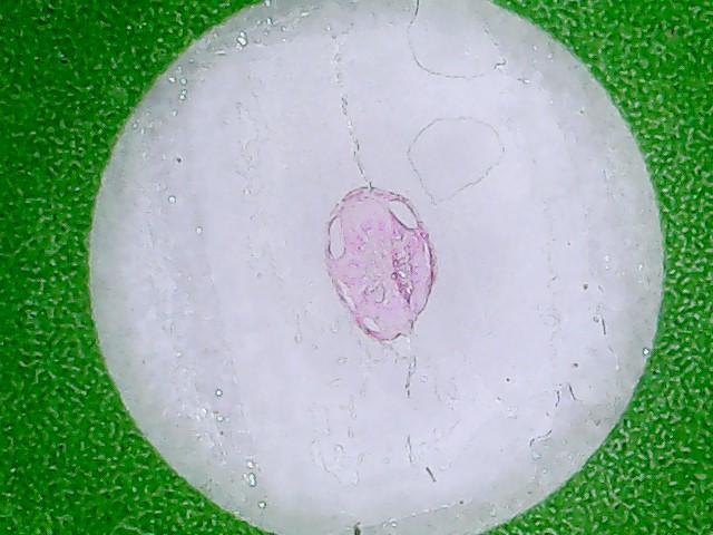
- Lotus Root: This slide sample shows the focused image of Lotus Root using the microscope.
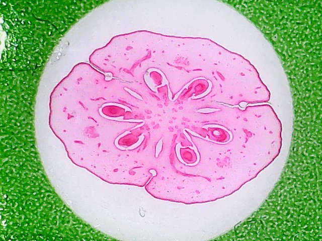
- Cucumber Ovary : This slide sample shows the focused image of Cucumber Ovary using the microscope.
- Onion epidermis: This slide sample shows the focused image of Onion epidermis using the microscope.
- Burdock Root: This slide sample shows the focused image of Burdock Root using the microscope.
- Celery Leaf: This slide sample shows the focused image of Celery Leaf using the microscope.
- Ginger Roots: This slide sample shows the focused image of Ginger Roots using the microscope.
- Cabbage Leaf: This slide sample shows the focused image of Cabbage Leaf using the microscope.
- Carrot root: This slide sample shows the focused image of Carrot root using the microscope.
- Corn Stem: This slide sample shows the focused image of Corn Stem using the microscope.